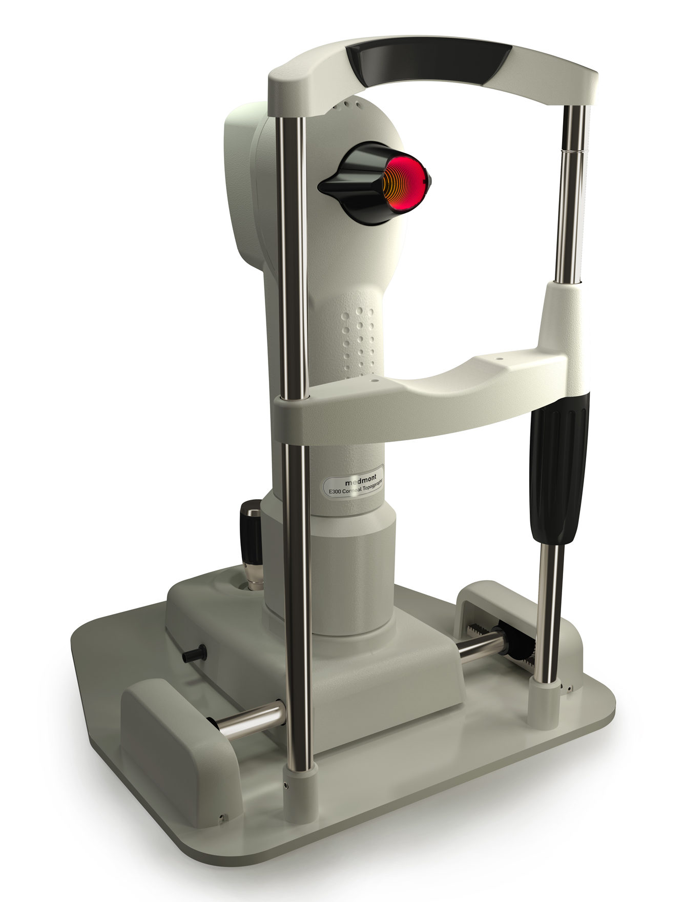
MEDMONT E300 CORNEAL TOPOGRAPHER
The Medmont E300 offers Extreme Accuracy for the Mapping of a Patients Cornea. It has a Standard Deviation of Error of less than 2 microns. To put that into perspective, the average human hair is 75 microns. It is also the only Placido Ring Topographer on the market that can map the cornea from Limbus to Limbus. This is a huge advantage when fitting Scleral or GP Lenses. Used extensively by the top Contact Lens Manufacturers and Optometry Universities throughout the world, the E300 is considered the Gold Standard for fitting Specialty Contact Lenses.
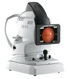
NIDEK NON-MYDRIATIC AUTO FUNDUS CAMERA
The NIDEK NON-MYDRIATIC FUNDUS CAMERA, AFC-330 is an ophthalmic camera that is indicated for use in capturing images of the retina and the anterior segment of the eye using a built-in color CCD camera without the use of mydriatic agent.
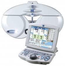
MARCO TRS-5100
The TRS-5100 replaces the manual refractor and allows the doctor to control the refraction process through a keypad. The TRS enhances the patient experience through a faster, more modern exam. The TRS-5100 generates extremely accurate and reliable refractions, minimizes staff variables, eliminates transcription errors, while smoothly integrating with EMR systems.
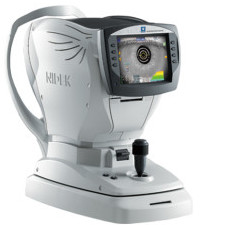
ARK-1 AUTOREFRACTOR / KERATOMETER
ARK-1 autorefractor/keratometer is equiped with the best technology today. The Super Luminescent Diode (SLD) makes it possible to accurately measure patients with cataracts, corneal opacities, IOLs, and post LASIK procedures. It also utilizes a multiple pupil zone imaging method combined with rotary prism technology, and auto alignment Eye Tracking System on the X-Y axes ensuring consistent, accurate, reliable and repeatable data.
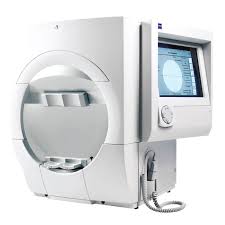
HUMPHREY VISUAL FIELD ANALYSER 750i
The Humphrey Visual Field Analyser 750i (HVFA), is a tool for measuring the human visual field, it is used by optometrists, orthoptists and ophthalmologists, particularly for detecting monocular visual field. The results of the Analyser identify the type of vision defect, and it provides information regarding the location of any disease processes or lesion(s) throughout the visual pathway. This guides and contributes to the diagnosis of the condition affecting the patient’s vision.